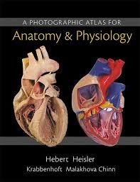
“A Photographic Atlas for Anatomy & Physiology (Looseleaf)” is an educational resource designed to complement studies in anatomy and physiology. This atlas is tailored for students in the health sciences and related fields, offering a comprehensive collection of high-quality photographs and illustrations that vividly depict the human body’s anatomical structures and physiological processes. The looseleaf format allows for easy handling and the possibility to only carry what’s needed for study or lab work, enhancing the learning experience.
The atlas includes detailed photographic representations of various body parts, systems, and functions, including skeletal, muscular, nervous, respiratory, circulatory, and digestive systems, among others. It serves as a visual guide to help students better understand the complex details of human anatomy and physiology, supporting their coursework, lab work, and preparations for exams.
Character Analysis
- Given its nature as an educational tool, the atlas does not contain narrative elements or characters but focuses on providing accurate and detailed visual information. It is designed to be an aid for both instructors and students, facilitating a deeper understanding of the subject matter through visual learning.
Themes and Analysis
- Visual Learning: The atlas emphasizes the importance of visual learning in the study of anatomy and physiology, offering clear and detailed images to aid in the comprehension of complex biological concepts.
- Anatomical Detail: It provides an in-depth look at the human body's anatomical structures, highlighting the intricacy and interconnectedness of different systems.
- Educational Utility: The resource is created with the educational needs of students and instructors in mind, offering a practical tool for enhancing classroom learning and independent study.
“A Photographic Atlas for Anatomy & Physiology (Looseleaf)” is a valuable educational resource for anyone studying or teaching anatomy and physiology. Its comprehensive visual content, combined with the flexibility of the looseleaf format, makes it an ideal companion for classroom instruction, lab sessions, and individual study. By offering detailed photographs and illustrations of the human body, the atlas helps demystify complex subjects, making them more accessible and understandable to students at various levels of their education. Whether used as a primary reference or a supplementary resource, this photographic atlas is an essential tool for enhancing the learning experience in the fields of health science and beyond.
If the summary caught your interest,
Consider reading the full book on AbeBooks.
Explore this book on AbeBooks
